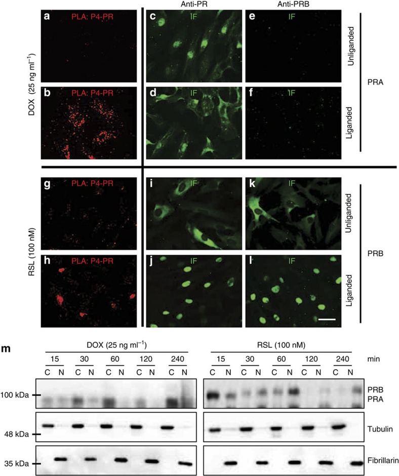
Figure 5. Liganded and unliganded PRA and PRB isoforms have differential cellular distribution.
Representative pictures of: in situ Proximity Ligation Assay (PLA) and immunofluorescence (IF) for total PRs and PRB in human myometrial cells with inducible PRA (a–f) and PRB (g–l) expression system. Cells were induced for 24 h with Dox (25 ng ml−1) for PRA and with RSL (100 nM) for PRB expression and then treated with vehicle or P4 (100 nM) for 2 h before subjecting to fixation and PLA analysis. Scale bar, 40 μm. (m) Cytoplasmic (C) and nuclear (N) fractionation analysis of PRA and PRB following stimulation with P4 for 15–240 min demonstrates gradual accumulation of PRB in the nucleus and PRA in the cytoplasm. Representative western blots and fractionation controls (Tubulin and Fibrillarin) are shown.

