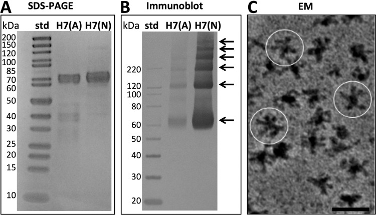
FIG 1.
Biochemical characterization of H7 hemagglutinin (HA). (A) SDS-PAGE analysis of purified HA proteins under denaturing and reducing conditions. The H7 proteins from the A/Anhui/01/2013 (H7N9) and A/Netherlands/219/2003 (H7N7) influenza viruses are designated H7(A) and H7(N), respectively. The first lane contains molecular mass standards (std); the second and third lanes contain purified HA proteins. (B) Immunoblot analysis of HA oligomerization under nondenaturing and nonreducing conditions. The first lane contains immunoblot standards; the second and third lanes contain HA proteins. Arrows indicate a ladder of at least six protein bands visible above background. (C) Image of a field of H7 (Netherlands) HA complexes revealed by negative-staining electron microscopy (EM) with a heavy metal stain, uranyl acetate. Image contrast is shown, with black areas representing proteins. Several complexes are circled. Bar, 50 nm.

