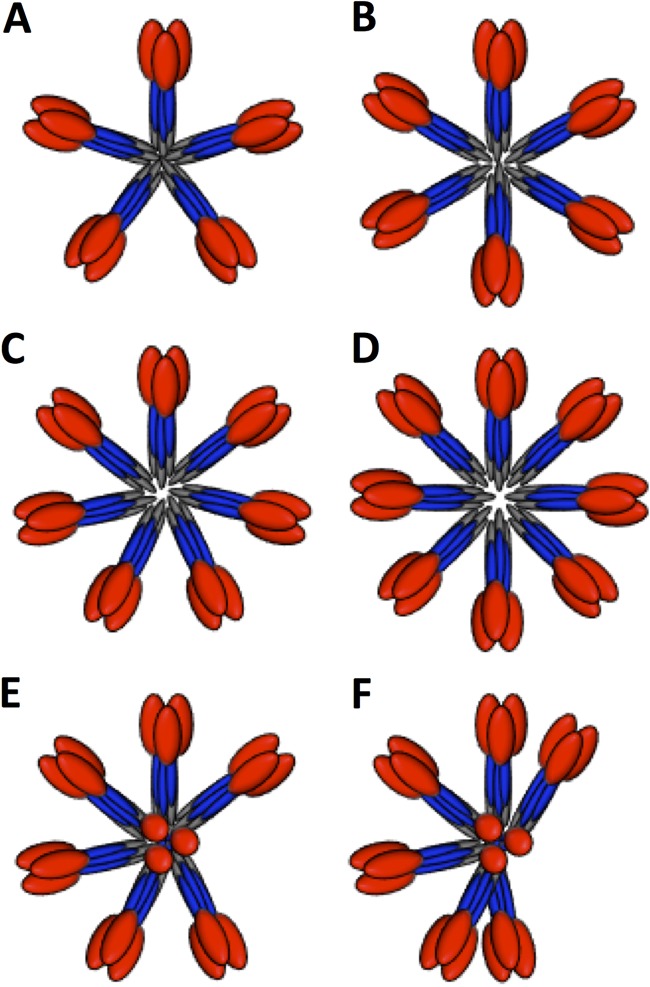
FIG 7.
Schematic models of HA complexes to illustrate the positional variations of constituent HA molecules. (A to D) Schematics of five, six, seven, and eight HA trimers, respectively, in planar starfish-like arrangements. (E) Schematic of seven HA trimers in an asymmetric starfish-like arrangement with one molecule in an upward-facing position. (F) Schematic of seven HA trimers in an asymmetric starfish-like arrangement with one molecule in an upward-facing position and various distances between lateral molecules. In panels E and F, one HA molecule is perpendicular in order to indicate a top axial view as opposed to lateral views. In all the schematic models, HA1 is shown in red, HA2 in blue, and transmembrane regions in gray.

