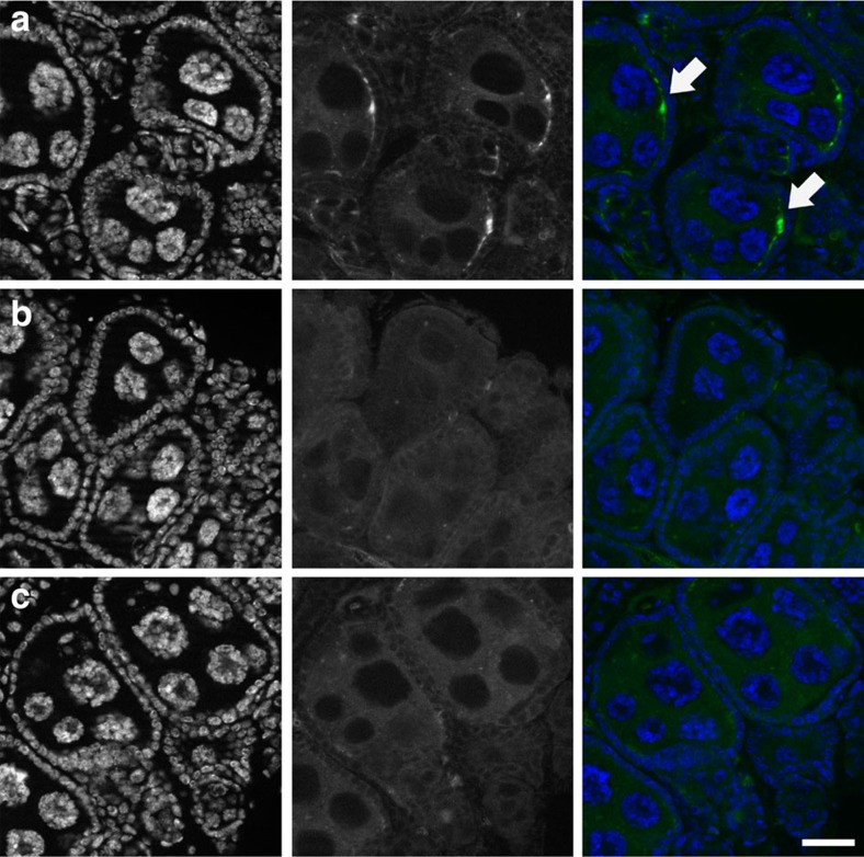
Figure 1. wAnga localizes to the female germline.
wAnga (in green, central panels) was visualized in the ovaries of 14- to 16-day-old A. coluzzii females by FISH. (a) wAnga is detected in ovarian follicles using a Cy3-labelled probe specific for 16S DNA (white arrows). (b,c) wAnga is absent from the follicles of tetracycline-treated control females (b) and in follicles of infected females in which the labelled probe was in competition with an identical unlabelled probe (1:20 labelled:unlabelled) (c). DNA is labelled with 4,6-diamidino-2-phenylindole (in blue, left panels). Scale bar, 20 μm.

