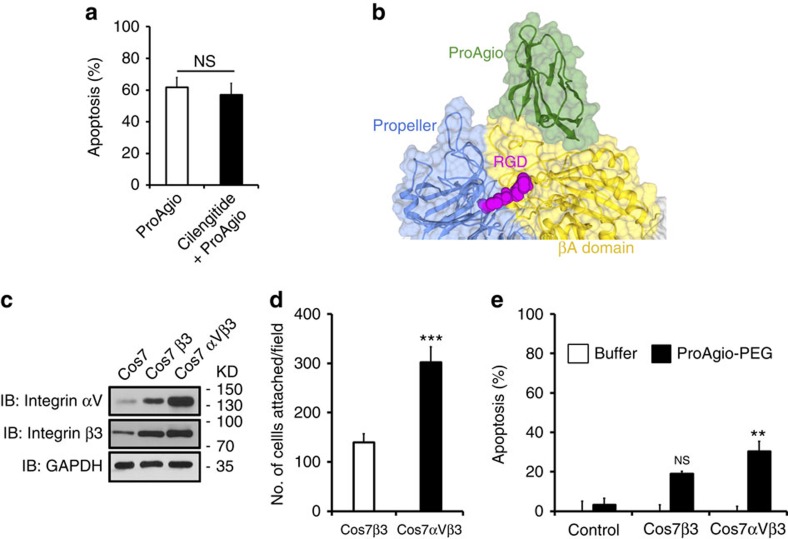
Figure 6. ProAgio and integrin αvβ3 interaction is independent of RGD binding.
(a) Apoptosis of HUVEC cells was measured by cell counting 24 h after treatment with 5 μM of ProAgio. The cells were incubated with 5 μM Cilengitide (filled bar) or buffer (open bar) before the ProAgio treatment. Cell apoptosis is presented as % apoptosis by defining the apoptosis of untreated cells as reference 0%. (b) Docking model of ProAgio (Green) with integrin αvβ3 in the presence of RGD (Red) using HADDOCK 1.2 program. (c) Levels of αv (IB: Integrin αV) and β3 (IB: Integrin β3) integrins in COS-7 cells with expression of β3 (COS7β3) or αvβ3 (COS7αVβ3) were analysed by immunoblot of cell lysates. Immunoblot of GAPDH (IB:GAPDH) is a loading control. (d) Cell attachment of COS-7 cells with expression of αvβ3 (COS7αVβ3) or β3 (COS7β3) to culture plate coated with ProAgio. The cell attachments are presented as total number of cells attached to the plate per view field (average of three fields). (e) Apoptosis of COS-7 cells with expression of αvβ3 (COS7αVβ3) or β3 (COS7β3) treated by 5 μM ProAgio. Cell apoptosis is presented as % apoptosis by defining the apoptosis of untreated cells as reference 0%. Error bars in a,d and e are s.d. from measurement of five independent experiments. NS P>0.05, **P<0.005, ***P<0.001 calculated by unpaired two-tailed Student's t-test. NS, not significant.

