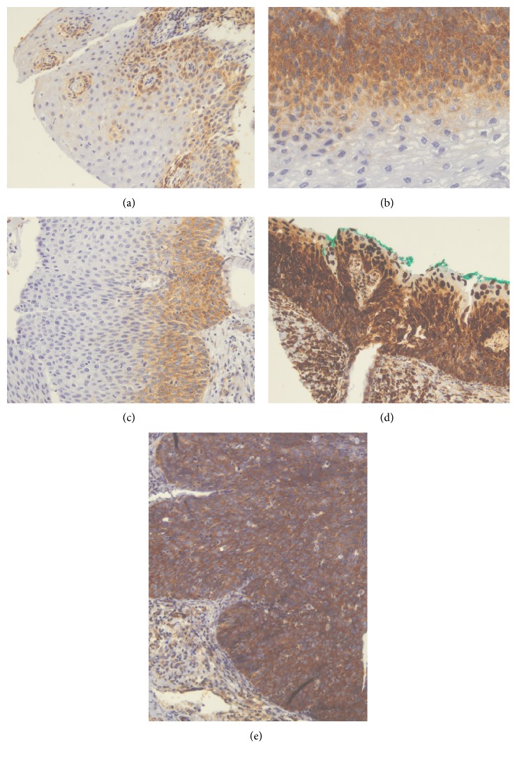
Figure 1.
Microphotographs showing the immunohistochemical expression of VEGF in cervical tissues. (a) Nonneoplastic squamous epithelium with weak expression (×100), (b) CIN 1 with moderate expression (×200), (c) CIN 2 with moderate expression (×100), (d) CIN 2 with strong expression (×100), and (e) CIN 3 with strong expression (×200).

