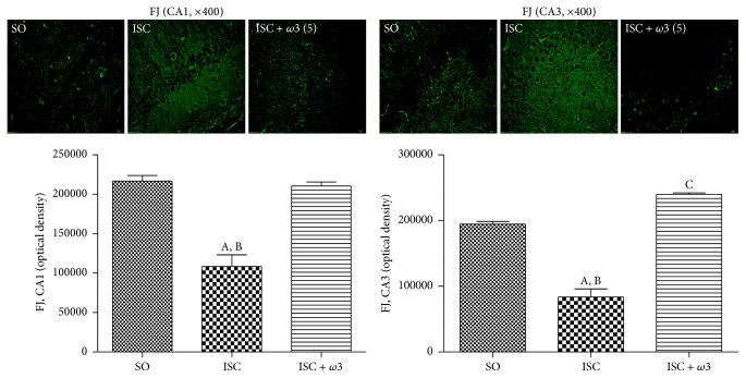
Figure 5.
ω3 treatments of ischemic animals (3 animals per group) drastically decreased the neuronal degeneration (visualized by an intense fluorescence to Fluoro-Jade) in hippocampus and cortex. The effects in CA1 and CA3 and DG are represented by photomicrographs and quantitative measurements performed with the Image J software, from 3 to 6 fields. The groups are sham-operated (SO); ischemic (ISC) untreated, and ischemic after ω3 treatments (5 mg/kg, for 7 days). CA1: (A) versus SO, q = 10.98∗∗∗; (B) versus ISC + ω3, q = 10.34∗∗∗. CA3: (A) versus SO, q = 14.57∗∗∗; (B) versus ISC + ω3, q = 20.55∗∗∗; (C) versus SO, q = 5.979∗∗ (one-way ANOVA and Newman-Keuls test as the post hoc test).

