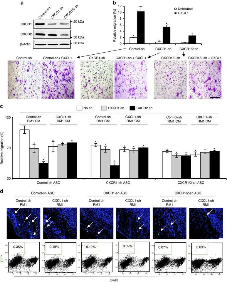
Figure 5. CXCR1/CXCR2 silencing inhibits ASC migration to tumours.
(a) Immortalized GFP-expressing mouse ASCs transduced with an untargeted shRNA (control-sh), or shRNA targeting either only CXCR1 (CXCR1-sh) or both CXCR1 and CXCR2 (CXCR1/2-sh) were subjected to immunoblotting with CXCR1 and CXCR2 antibodies. Experiment was performed twice with similar results; representative scans are shown. (b) Cells from a were subjected to transwell chamber migration through 8-μm pores toward medium with or without 5 ng ml−1 CXCL1. Plotted are relative migrated cell numbers normalized to migration of SC ASCs towards 10% FBS (100%). Shown below are representative bright-field micrographs of crystal violet-stained transwell membranes corresponding to each treatment group. Scale bar, 100 μm. (c) Cells from a were subjected to transwell chamber migration through 8-μm pores towards CM from control-shRNA or CXCL1-shRNA RM1 cells supplemented with antibodies (abs) against CXCR1 or CXCR2 where indicated. (d) Representative sections of tumours formed by control-sh-RM1 and CXCL1-sh RM1 cells grafted into the nude mice (n=6) that received subcutaneous contra-lateral injections of control-sh, CXCR1-sh or CXCR1/2-sh GFP-expressing ASCs. Arrows indicate tumour-engrafted ASCs (green), recruitment of which is reduced by both CXCL1 and CXCR1 silencing. Scale bar, 100 μm. Nuclei are blue. Representative flow cytometric analyses of suspended tumour cells enumerating injected ASCs (GFP+) among viable (4,6-diamidino-2-phenylindole (DAPI) negative) cells. Note ASC frequency reduction by CXCL1 and CXCR1 silencing. For b–d, experiments were performed in technical triplicate. Graphs show mean±s.e.m.; *P<0.05 (Student's t-test) versus control-sh CM.

