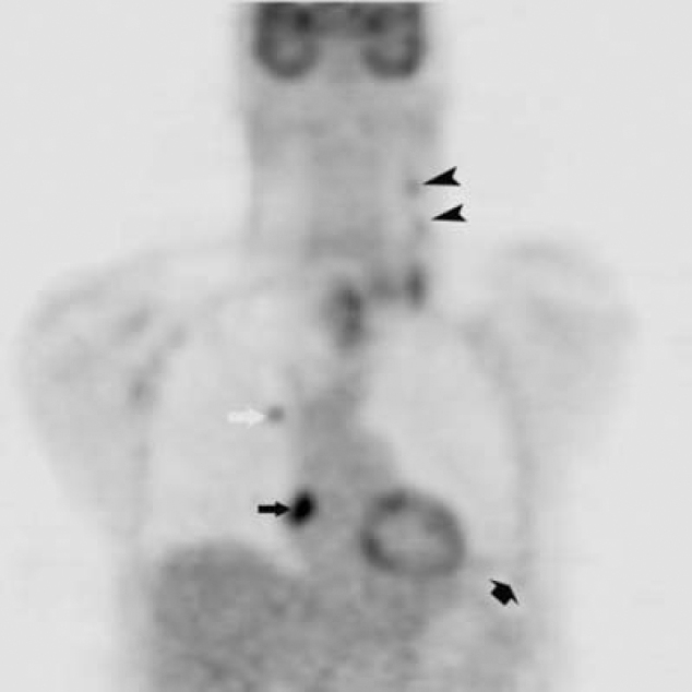
Figure 1D.

47-year-old man with metastatic head and neck cancer. Coronal reformatted PET corresponding to Fig. 1C shows a right atrial mass (black arrow)(SUV max = 10.2), right upper lobe pulmonary nodule (white arrow) (SUV max = 4.0), left cervical nodal disease (black arrowheads), and left lower lobe infarct (short black arrow) (SUV max = 2.1).
