. 2016 Jun 1;28(2):165–171. doi: 10.5792/ksrr.2016.28.2.165

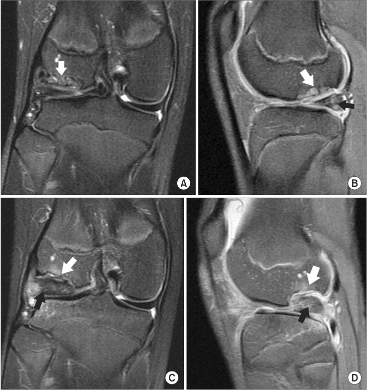
Fig. 3. Initial preoperative magnetic resonance imaging (MRI) performed at another hospital showing the osteochondral lesion with subchondral cysts (white arrow) in the posterior aspect of the right lateral femoral condyle (LFC) in coronal plane (A) and the osteochondral lesion (white arrow) with posteriorly displaced lateral discoid meniscus (black arrow) in the posterolateral aspect in sagittal plane (B). Follow-up (5 months after initial MRI) preoperative MRI performed at our hospital showing the more progressed osteochondral lesion (white arrow) in coronal plane (C) and the massive defect of the LFC (white arrow) with retorn lateral meniscus adhering to the LFC (black arrow) in sagittal plane (D).

Fig. 3