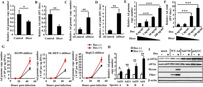
Figure 4. Dicer-mediated suppression of Ad replication.
(A–D) HeLa cells were transfected with a Dicer-expressing plasmid (p3XFLAG-CMV10-Dicer) (A,B) or siRNAs (C,D), followed by infection with WT-Ad at an MOI of 5. After 24 h incubation, the copy numbers of WT-Ad genomic DNA (A,C) and the IFU titers of progeny WT-Ad (B,D) in the cells were determined by real-time PCR analysis and infectious titer assay, respectively. (E,F) HeLa-shDicer cells were cultured in Dox-free or Dox-containing medium at the indicated concentrations for 48 h, followed by infection with WT-Ad at an MOI of 5. After 24 h incubation, the copy numbers of WT-Ad genomic DNA (E) and IFU titers of progeny WT-Ad (F) in the cells were similarly determined. (G) H1299-shDicer, SK HEP-1-shDicer, and HepG2-shDicer cells were cultured in Dox-free or Dox-containing (100 ng ml−1) medium for 48 h, followed by infection with WT-Ad at an MOI of 5. At the indicated time point, copy numbers of WT-Ad genomic DNA were determined by real-time PCR analysis. (H) HeLa-shDicer cells were cultured in Dox-free or Dox-containing (100 ng ml−1) medium for 48 h, followed by infection with Ad31, Ad11, Ad35, or Ad4 at 100 virus particles (VP) per cell. After 24 h incubation, the copy numbers of each Ad genomic DNA were determined by real-time PCR analysis. These data (A–H) are expressed as the means ± S.D. (n = 3–4). (I) Phosphorylated eIF2α protein levels following infection with WT-Ad or Sub720 in HeLa-shDicer cells were analyzed by western blotting analysis. The cells were cultured in Dox-free or Dox-containing (100 ng ml−1) medium for 48 h, followed by infection with WT-Ad or Sub720 at an MOI of 5 for 24 h. HeLa-shDicer cells cultured in Dox-free or Dox-containing (100 ng ml−1) medium for 48 h were transfected with polyI:C (1 μg ml−1), followed by western blotting analysis after 6 h incubation. Hexon and fiber proteins are major Ad capsid proteins. *p < 0.05, **p < 0.01, ***p < 0.001 (Student’s t-test).

