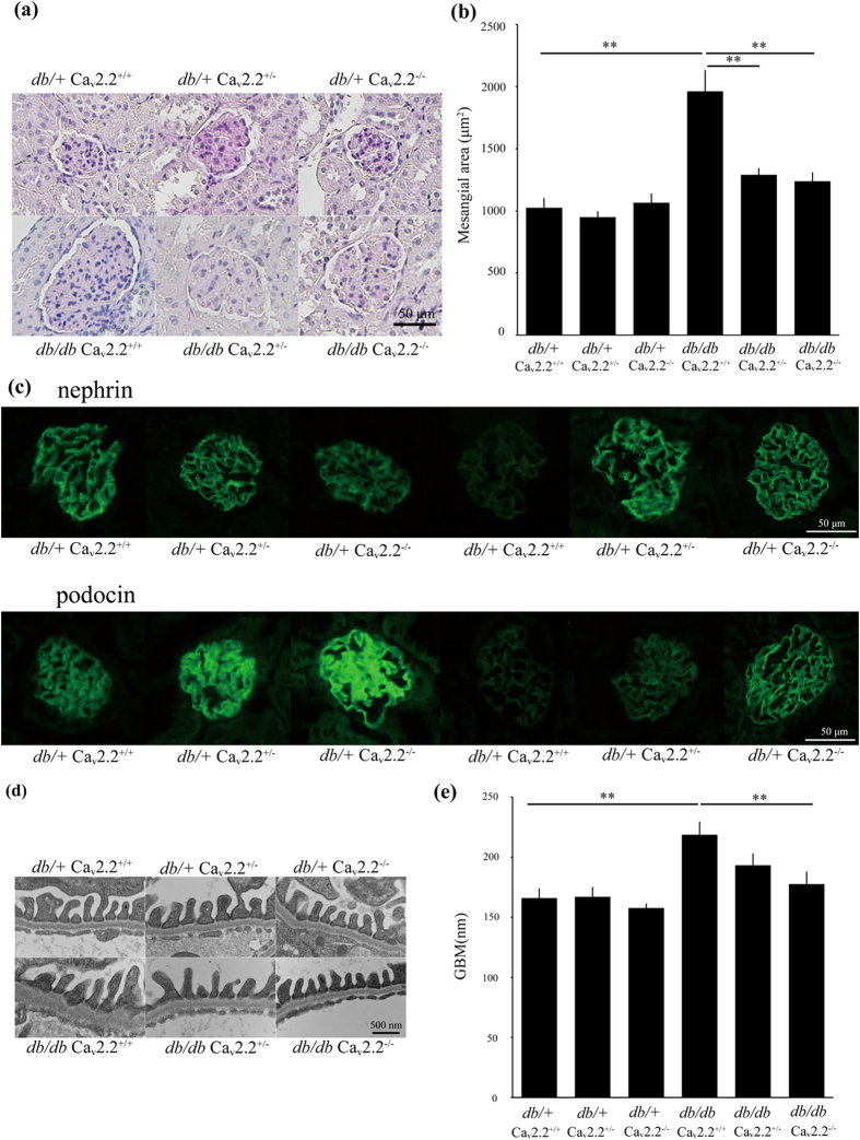
Figure 4. Histologic examination of a glomerulus.
(a) Light microscopic analyses were performed at 16 weeks of age, stained with periodic acid-Schiff. db/db Cav2.2−/− mice showed reduced mesangial expansion compared with db/db Cav2.2+/+ mice. Scale bar = 50 μm. (b) Mesangial area in a glomerulus at 16 weeks of age. Mesangial area was increased in db/db Cav2.2+/+ mice and was suppressed in db/db Cav2.2−/− mice. (c) Immunostaining for nephrin and podocin. db/db Cav2.2−/− mice maintained of nephrin and podocin to the same level with db/+ mice. Scale bar = 50 μm. (d,e) Electron microscopic analyses of glomeruli of experimental mice at 16 weeks of age. GBM thickness was ameliorated in db/db Cav2.2−/− mice. Scale bar = 500 nm. **p < 0.01, vs. db/db Cav2.2+/+ mice.

