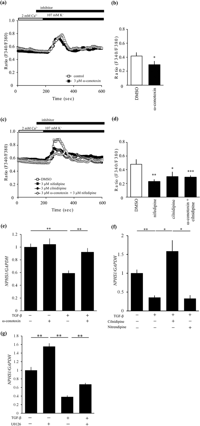
Figure 7. Expression of N-type calcium channels in cultured human podocytes and the functional role of these channels in depolarization-induced Ca2+ response.

(a,b) Inhibitory effect of 3 μM ω-conotoxin (black squares) on depolarization-induced Ca2+ responses in human podocyte (n = 27–39) compared with control (white squares). (a) Averaged time courses of depolarization-induced Ca2+ response in human podocyte. (b) The bars represent the differences between the maximum of ration and the value of steady state. *p < 0.05 vs. control. (c,d) Inhibitory effect of 3 μM cilnidipine and 3 μM nifedipine on depolarization-induced Ca2+ responses in human podocyte (n = 20–30). (c) Averaged time courses of depolarization-induced Ca2+ response in human podocyte. 3 μM nifedipine; black squares, 3 μM cilnidipine; black circles, 3 μM ω-conotoxin + 3 μM nifedipine; white circles, DMSO; white squares. (d) The bars represent the differences between the maximum of ratio and the value of steady state. *p < 0.05, **p < 0.01, ***p < 0.001 vs. DMSO. (e) TGF-β1 (20 ng/ml) suppressed NPHS1 expression in cultured human podocytes. Its inhibition was canceled by 100 nM ω-conotoxin. n = 11 for TGF-β (−) groups and n = 12 for TGF-β (+) groups. (f) Cilnidipine (10 μM) not nitrendipine (10 μM) ameliorated reduction of NPHS1 expression induced by 5 ng/ml TGF-β1 in podocytes. Vehicle (n = 5) and other groups (n = 6, each). *p < 0.05, **p < 0.01. GAPDH was used as control.
