
Figure 1.
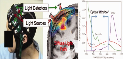
Functional near‐infrared spectroscopy (fNIRS) is used to noninvasively measure changes in oxy‐ and deoxyhemoglobin in the brain. A grid of fiber optic‐based light sources and detectors is mounted into a flexible head cap worn by the participant (left). Each of these source‐detector pairs measures light from a diffuse volume of tissue beneath the pair (center). This light can reach ∼ 5–8 mm into the cortex at a source‐detector spacing of 3.2 cm. [Color figure can be viewed in the online issue, which is available at http://wileyonlinelibrary.com.]