
Figure 4.
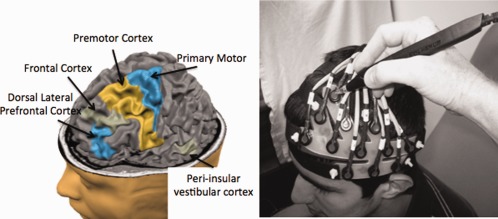
Based on previous fMRI studies, the left dorsal–lateral prefrontal cortex (DLPFC; BA 46), superior and inferior frontal cortex, inferior temporal gyrus (peri‐insular vestibular cortex; PIVC), premotor (BA 6), and primary (BA 4), and secondary motor cortices were targeted. The fNIRS sensors were mounted into a cap (shown without the neoprene hood for clarity on the right; also see Fig 1). A three‐dimensional wand (Polhemus) was used to mark the location of the sensors for later registration to structural MRI information. The image above (left) was generated using the BrainVoyager tutorial (http://www.brainvoyager.com). [Color figure can be viewed in the online issue, which is available at http://wileyonlinelibrary.com.]