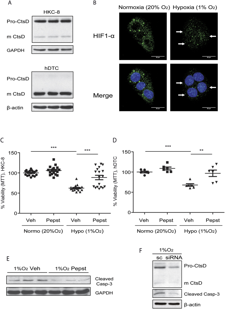
Figure 6. Pepstatin A reduces hypoxic induced apoptosis in tubular epithelial cells.
CtsD and GAPDH WB of HKC-8 cells and CtsD and β-actin WB of hDTC. (A) HIF-1α immunostaining in HKC-8 cells under normoxic (20% O2/5% CO2) or hypoxic (1% O2/5% CO2) conditions for 48 hours. (B) White arrows point to HIF-1α located within the nuclei. Percentage of metabolically active viable cells assessed by MTT assay in HKC-8 cells (C) or hDTC passage 2 (D) treated with vehicle or Pepstatin A under normoxic (20% O2/5% CO2) or hypoxic (1% O2/5% CO2) conditions for 48 hours. Cleaved caspase-3 and GAPDH WB in HKC-8 cells under hypoxic conditions for 48 hours treated with vehicle or Pepstatin A. (E) Cleaved caspase-3 and β-actin WB in HKC-8 cells under hypoxic conditions for 48 hours treated with scramble or siRNA against CtsD. (F) N = 3, 1 way ANOVA,*P ≤ 0.05 or **P ≤ 0.01.

