Fig. 3.

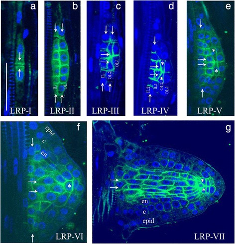
Changes in PIN1 expression during lateral root primordium development. a–g The seven stages of primordium development are shown in roman numerals. IL (IL1, IL2)—inner layers, OL (OL1, OL2)—outer layers. Pre-specified QC cells are marked by asterisks. Developing tissues: epid—epidermis, c—cortex, en—endodermis. White arrows show the directions of auxin flux. Anti-PIN1 staining is in green, DAPI is in the blue channel. Bars = 50 μm
