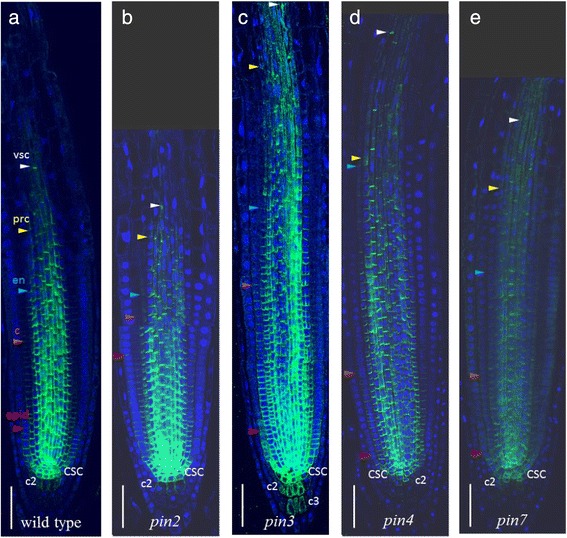
Fig. 6.

PIN1 expression patterns in pin mutants (b – e) compared to wild type (a). CSC—columella stem cell, c2—the second columella tier, c3—the third columella tier, epid—epidermis, c—cortex, en—endodermis, prc—pericycle, vsc—vasculature. Coloured triangles—the end of the expression domain in the respective layer. Anti-PIN1 staining is in green, DAPI is in the blue channel. Bars = 50 μm
