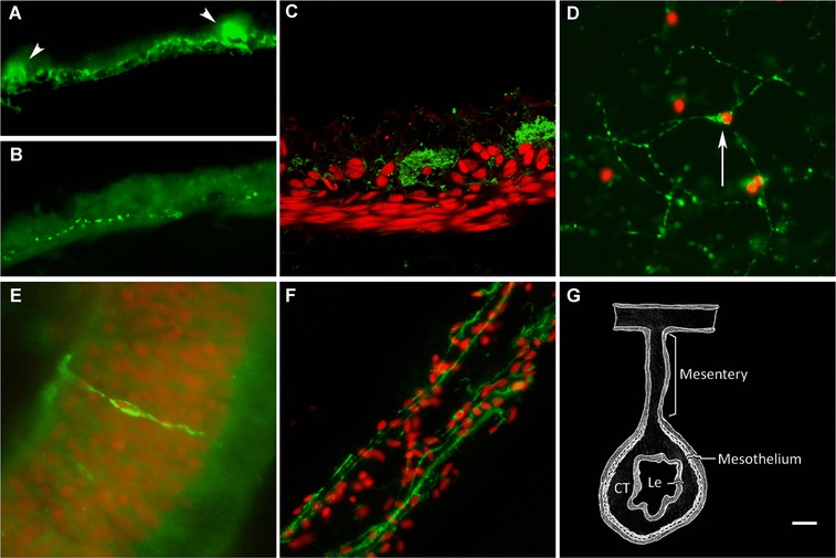
Figure 1.

Subdivisions of the holothurian ENS. (A) Mesothelial plexus labeled with monoclonal antibody RN1 showing an extensive fiber plexus and fiber bundles (arrowhead) while (B) other markers (anti‐PH3) only labeled discrete fiber populations. (C) Confocal image showing RN1 label fiber (green) distribution in relation to the muscle layer (labeled with Cy3‐labeled phalloidin, red). (D) Connective tissue plexus as labeled by RN1. Arrow points to a neuron‐type cell. (E) Neuroendocrine cell labeled with RN1 in the luminal epithelium. (F) RN1 labeling of fibers in the mesothelial layer of the mesentery. (G) Diagram showing the intestinal tissue layers. CT, connective tissue layer; Le, luminal epithelial layer. Nuclei are stained with DAPI (red) in (D), (E), (F). Bar (A, B) 10 μm; (C, E, F) 20 μm.
