
Figure 9.
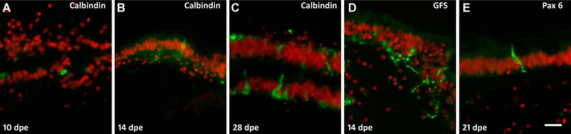
Regeneration of neuroendocrine cells. (A) Initial appearance of calbindin‐immunoreactive cells in a 10 dpe luminal epithelium is observed soon after lumen formation. At 14 dpe (B) calbindin‐immunoreactive cells acquire neuroendocrine morphology and by 28 dpe (C) the number of cells has increased significantly (the figure shows two adjacent luminal epithelia). GFS‐immunoreactive cells (D) also appear during the second week of regeneration showing their extended fiber connections. (E) Pax6‐immunoreactive neuroendocrine cells appear at 21 dpe. Bar 20 μm.