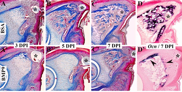
Figure 3.

BMP9 treatment delays ossification during regeneration (top is dorsal and proximal is to the left). The microcarrier bead associated with the treatment is indicated with an asterisk. (A)−(D) BSA treated control digits. (A′)−(D′) BMP9 treated digits. (A)−(C), (A′)−(C′) Mallory's triple stained histological sections of amputated digit tips. By 3 DPI the difference between control regenerates (A) and BMP9 treated digit amputations (A′) is subtle. Both accumulate blastema cells distally but ossification of BMP9 treated digit stumps appears to be delayed. By 5 DPI ossification is prominent in the stump and proximal region of control regenerates (B) whereas ossification is clearly delayed following BMP9 treatment (B′). By 7 DPI ossification is occurring throughout the control regenerate (C) whereas ossification is restricted to the stump region of BMP9 treated digits (C′). In situ hybridization to localize transcripts for the osteogenic marker gene, Osteocalcin (Ocn), validate the histological results. Ocn is expressed throughout the control regenerate at 7 DPI (D) whereas transcripts are localized to the periphery (arrowheads) of the stump in BMP9 treated digits (D′).
