
Figure 5.
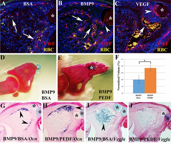
(A)−(C) Revascularization based on immunohistochemical analysis for VWF (red) 7 days after microcarrier bead (*) implantation. Sections were counterstained with DAPI, red blood cells (RBC, yellow) are autofluorescent, distal is to the right. (A) Staining of control BSA treated digits show blood vessels in the periphery of the digit stump (arrows) and sparse staining in the blastema. (B) VWF staining of BMP9 treated digits show blood vessels throughout the digit stump (arrows) and extending into the blastema (arrowhead). (C) After VEGF treatment an extensive vascular response is evident in the digit stump (arrow) and in blastema cells (arrowhead) associated with the bead (*). (D)−(F) The inhibitor activity of BMP9 is rescued following treatment with the anti‐angiogenic factor PEDF. (D) Whole mount skeletal staining shows that the BMP9 inhibited regenerative response is not modified by a control BSA bead implanted 1 day later. (E) BMP9 treatment followed by the implantation of a PEDF bead 1 day later restores the regenerative ability as shown by whole mount skeletal staining of digits at 14 DPI. (F) Bone volume analyzed by microCT is significantly increased by PEDF treatment. Data are normalized to the BMP9 inhibited BSA control digits, P < 0.01 (*). (G)−(J) In situ hybridization of control and PEDF rescued regenerates 7 days after implantation. (G) BMP9 inhibited Ocn expression (arrowheads) is not modified by BSA control bead implantation. (H) In PEDF rescued BMP9 inhibited digits Ocn expression extends distally (arrowhead) into the blastema region. (I) Upregulation of Vegfa transcripts (arrowhead) in the stump of BMP9 inhibited digits is not modified by control BSA treatment. (J) In PEDF rescued BMP9 inhibited digits Vegfa expression is downregulated and appears similar to untreated regenerates at a similar stage (see Fig. 1C).