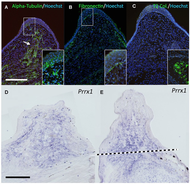
Figure 2.

Gene expression pattern in the bump. (A) Neural tubulin (acetylated alpha‐tubulin) was visualized by immunofluorescence. The white arrow indicates the thick nerve bundle observed in the center of the bump. The deviated nerves abundantly penetrate into the overlying epithelium (inset). (B) One of the classical blastema marker genes, fibronectin, was investigated. The signal of fibronectin can be seen in the blastemal region and the epithelium−mesenchyme boundary shows intense fibronectin signal (inset). (C) Type II collagen expression. Type II collagen is a cartilage marker gene and negative in the bump. The inset shows the signal in the limb cartilage located in the proximal region as the positive control. (D, E) Prrx1 expression. Prrx1 can be seen in the blastemal mesenchyme in the ALM blastema (D) and the regular blastema in the amputated limb (E). The dotted line in (E) indicates the presumptive amputation plane. All conditions of the in situ hybridization were the same in (D) and (E). Scale bars in (A) and (D) are 0.5 mm.
