Figure 4.

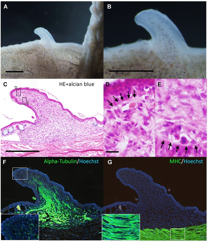
The induced bump did not contain cartilaginous cells. (A, B) Dorsal views of the day 20 bump. The induced bump stopped growing at day 20. (C) Histology of the bump. There are no Alcian‐blue‐positive cartilage cells in the mesenchymal region. (D, E) Higher magnification of the boxed regions in (C). Arrows in (D) and (E) indicate developing basal laminae. (F) Axons were visualized by immunofluorescence using anti‐acetylated alpha‐tubulin. In the distal region, highly innervated axon fibers in the epithelium were not observed in the day 20 bump (inset). (G) Myosin heavy chain (MHC) expression. There were no MHC‐positive myogenic cells in the bump. Scale bars in (A) and (B) are 1 mm. The scale bar in (C) is 500 μm. (C), (F), and (G) are the same magnification. The scale bar in (D) is 20 μm. (D) and (E) are the same magnification.
