
Figure 3.
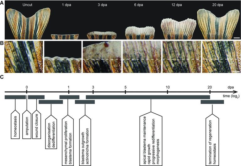
The regeneration process of the caudal fin in zebrafish. (A) Time‐lapse imaging of the same fin during the regeneration process at 27°C. Uncut, the original fin prior to amputation presents a bi‐lobed morphology. At 1 dpa, white tissue above the amputation consists of the wound epidermis and a few blastema cells. At 3 dpa, a white excrescence above the amputation plane contains the blastema, which, despite its uniform appearance, exhibits subdivisions at the cellular and molecular level. At 6 dpa, the outgrowth extends very rapidly; the white tissue is maintained at the fin margin, while the proximal outgrowth starts to display bone structures and pigmentation, which are the macroscopic markers of tissue redifferentiation. At 12 dpa, fin regeneration is at its advanced stage. At 20 dpa, the size of the fin nearly reaches its original size and pattern. The white margin of tissue remains at the tip for homeostatic growth/regeneration. (B) Higher magnifications of the fin surface at the position of amputation (white dashed line) at the respective time points are indicated in the upper panel (A). (C) The milestones of the fin regeneration process. Scale bars: (A) 1000 μm; (B) 200 μm.