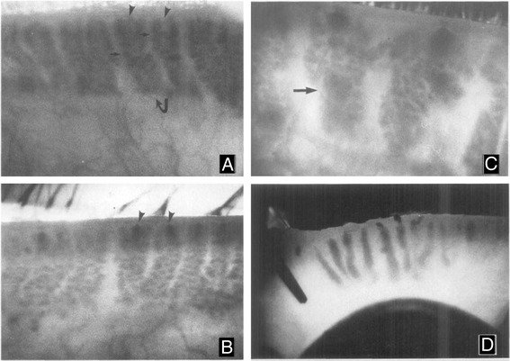
Fig. 1.

Meibography of the rabbit eyelids from normal (a), and 2 % epinephrine treated eyes for 1–2 months (b), 3–4 months (c) and 6 months (d). Normal glands (a. curved arrow) appear as grape-like clusters of individual acini (small arrow) with indistinct gland orifices (a, arrowheads). After 1–2 months of treatment, the orifices of the glands become detectible as dark spots at the leading edge of the glands (b, arrowheads). Progression of hyperkeratinization into the gland is detected by 3–4 months after treatment as large, dark cystic structure that obscure the normal acini (c, arrows). After six months of treatment the meibomian glands are replaced by large, dense keratic cysts (d). Magnification A-C: 24x and D: 10x
