
Figure 2.
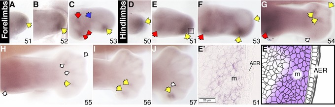
Sulf2 expression in X. laevis stage 51−53 forelimbs (A−C) and stage 50−57 hindlimbs (D−J) examined by a whole mount in situ hybridization. Specific staining is dark purple. Red arrows in the more proximal part indicate gene expression at the putative shoulder/hip and elbow/knee joints; blue arrows indicate expression at the future wrist/ankle joints; yellow arrows indicate transcript expression in the autopod region; empty arrows indicate reduced level of expression. Roman numerals indicate digit number. The boxed region in E indicates area of histological section shown in E′, accompanied by the corresponding schematic (E″) showing area of gene expression in the mesenchyme. All limbs are oriented proximal to the left and posterior uppermost, with the tadpoles lying on their right side. AER, apical epidermal ridge; m, mesenchyme.