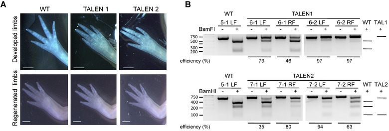
Figure 2.

TALEN‐targeted animals show edited tsp‐1 locus in the limbs and normal limb morphology. (A) Morphology of developed limbs and regenerated limbs 6 weeks post‐amputation from wild‐type and TALEN‐targeted animals. Scale bar indicates 1 mm. (B) PCR amplicon of the tsp‐1 locus from limbs from wild‐type and TALEN‐injected juvenile animals digested by restriction enzyme indicated edited tsp‐1 locus. Genomic DNA from one forelimb of WT (wild‐type; non‐injected) or TALEN mRNA‐injected animals was used in each lane. For each sample, an equal amount of PCR product, not incubated with the restriction enzyme, was loaded as an undigested control. The predicted patterns of DNA fragments after restriction enzyme digestion are illustrated on the right. The first lane from the left is the DNA ladder. PCR amplicons from TALEN1‐ and TALEN2‐mRNA‐injected embryos were cleaved by BsmFI or BamHI, respectively.
