Figure 3.

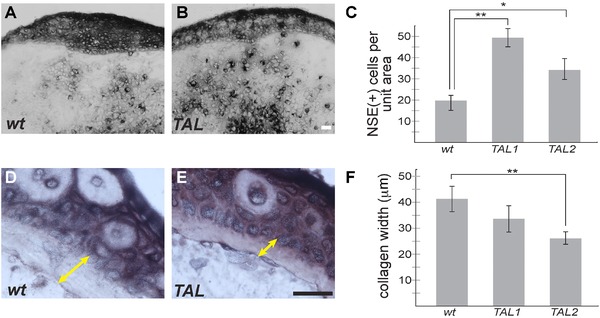
TALEN‐targeted animals show increased macrophage and monocyte infiltration in regenerating limbs and decreased stump collagen deposition. NSE staining was performed to detect monocytes and macrophages in regenerating juvenile limbs at 6 days post‐amputation. (A) Wild‐type sibling control. (B) TALEN‐targeted tsp‐1 deletion animal. (C) The quantification of NSE positive cells (A, B, black) within the blastema mesenchyme were quantified (N = 14, 6, 16 limbs for wt, TAL1 and TAL2 respectively). (D), (E) Subepidermal collagen thickness was measured (yellow double arrow) in control and TALEN‐targeted stumps, and quantified in (F) (N = 14 controls; 6 TAL1; 16 TAL2). Scale bars in all images are 50 μm; *P < 0.05, **P < 0.01; error bars indicate SEM.
