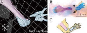
Figure 2.

Morphology of the radius and ulna regenerated without interaction with the remaining humerus. (A) 3D image constructed using EFIC image of the regenerated skeletal elements after joint and humerus amputation. The remaining tissues are segmented in pink, and the regenerated tissues are segmented in blue. (B) Whole‐mount bone and cartilage staining of the regenerated skeletal elements after joint and humerus amputation. Bones are stained magenta, and cartilage is stained blue. The radius and ulna were regenerated without interacting with the remaining humerus, and in this case the proximal structures of the radius and ulna were not completely regenerated (arrowheads), as shown (C) in a schematic drawing.
