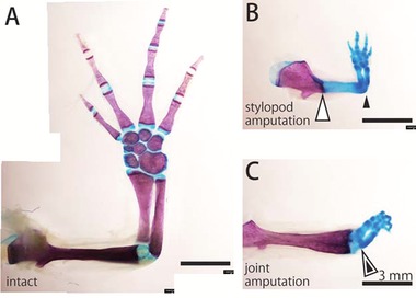
Figure 4.

Skeletal structure of the regenerated limb. Whole‐mount bone and cartilage staining of (A) the intact forelimb, (B) the regenerated forelimb at 70 days after stylopod amputation, and (C) the regenerated forelimb at 70 days after joint amputation. Bones are stained magenta, and cartilage is stained blue. White arrowheads indicate the amputated site and black arrowheads indicate the regenerated elbow.
