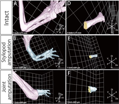
Figure 5.

3D reconstruction image of bone and cartilage obtained by EFIC. The 3D reconstructed image of the skeletal elements in (A) the intact forelimb, (B) the regenerated forelimb at 70 days after stylopod amputation, and (C) the regenerated forelimb at 70 days after joint amputation. The remaining tissues are segmented in pink and the regenerated tissues are segmented in blue. We measured the volume of (D) the radius of the intact forelimb, (E) the radius at 70 days after stylopod amputation, and (F) the radius at 70 days after joint amputation. The joint surfaces of the radius whose areas were measured are segmented in yellow in (D), (E), and (F).
