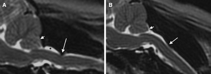
Figure 2.

T2‐weighted sagittal MR images of the craniocervical junction in a cavalier King Charles spaniels positioned in an extended/straight position (A) and in a flexion, resembling a standing posture (B). Dorsal atlantoaxial (AA) band‐associated compression (long arrow) is more prominent in flexion than in extension. A Chiari‐like malformation is also present (short arrow), along with dorsal subarachnoid space dilation cranial to the AA band (*).
