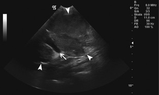
Figure 1.

Longitudinal gray‐scale ultrasonographic image: An ovoid slightly heterogenous lobulated mass of low echogenicity (white arrowheads) containing a large vessel (white arrow) is visible. The cranial and caudal extent of the lesion is not included in the images.
