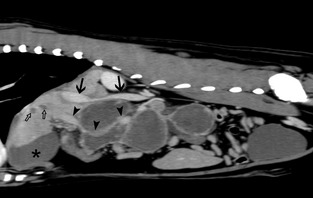
Figure 2.

Postcontrast sagittal computed tomographic images of the abdominal cavity, soft‐tissue algorithm. The sagittally reformatted image is slightly to the right of the midline: Note the ring enhancement of the lesion while the center noncontrast enhancing and hypoattenuating (Mean 20 HU) indicating central necrosis or exudate. The caudal vena cava (arrows) is narrowed at the level of the cranial aspect of the space‐occupying lesion. Ventral displacement of the portal vein (arrowheads) caudal to the liver hilum is present. Several ovoid hypoattenuating noncontrast enhancing areas are visible within the liver parenchyma (open arrows). The asterisk (*) indicates the gallbladder.
