Figure 3.

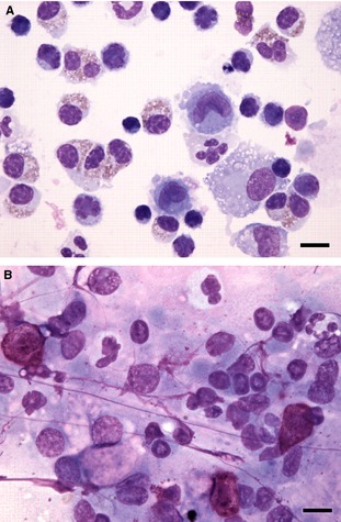
BALF cytospin cytology (A) and tracheal brush cytology (B) from a dog. There is discordance in the types of inflammation present. The BALF cytology has increased numbers of eosinophils and small mature lymphocytes, indicative of eosinophilic and lymphocytic inflammation. The tracheal brush cytology has increased numbers of mast cells (upper left, bottom right) and increased numbers of neutrophils (upper left, center and right), indicative of mastocytic and neutrophilic inflammation. There was also eosinophilic inflammation elsewhere in the brush cytology but lymphocytic inflammation was not detected. Note the brush cytology sample has some streaming nuclear material that is common in this sample type. Both samples also contain some background mucus and some lysed cells. 60× objective magnification, Wright Giemsa stain. Bar = 10 μm.
