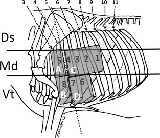
Figure 1.

Thoracic ultrasonographic and auscultation sites used in preweaned calves. Sites 1–8: Ultrasonographic sites at which thoracic examination was performed systematically. The median (Md) to ventral (Vt) parts of the thorax were divided into 4 auscultation areas (A–D). The auscultation findings were compared with ultrasonographic findings. The dorsal (Ds) third of the thorax was not examined.
