
Figure 2.
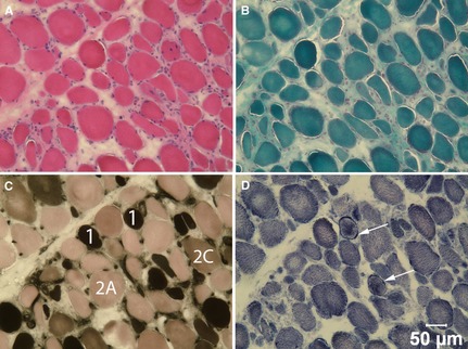
Cryosections from the biceps femoris muscle were stained with H&E (A) and modified Gomori trichrome (B), and reacted with myofibrillar ATPase at pH 4.3 (C) and NADH‐TR (D). Type 1, 2A and 2C fibers are labeled in image c and arrows denote sarcolemmal deposits of NADH‐TR positive material in d. Bar = 50 μm for a–d.