
Figure 4.
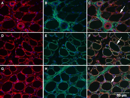
Cryosections from the biceps femoris muscle of the Labrador Retriever with sarcolemmal‐specific collagen VI deficiency (top and middle rows) and a dystrophin‐deficient dog (bottom row) were incubated with monoclonal or polyclonal antibodies against collagen IV (A,D,G), collagen VI (B,H), laminin α2 (E), and merged (C,F,I). Absence of sarcolemmal staining with the antibody against collagen VI was evident in the dog with sarcolemmal‐specific collagen VI deficiency (C, merge, arrow points to red sarcolemmal staining for only collagen IV), but present in the sections stained for laminin α2 (F, merge, arrow points to yellow sarcolemmal staining) and from the dystrophin‐deficient muscle (I, merge, arrow points to yellow sacolemmal staining). Bar = 50 μm for all images.