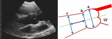
Figure 2.

Left parasternal cranial long‐axis view of the aortic root and schematic drawing of aortic root measurements at end‐diastole: (a) Valsalva sinus diameter; (b) sinotubular junction diameter; (c) ascending aortic diameter. The (c) diameter is measured at a distance from the sinotubular junction equal to (b).
