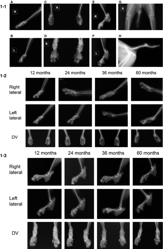
Figure 1.

Radiographs of the limbs and tail of the cat in case 1. 1–1 Radiographs at initial diagnosis (0 month). (A) Right carpus (lateral). (B) Left carpus (lateral). (C) Both carpi (dorsoventral, DV). (D) Both tarsi (DV). (E) Right tarsus (lateral). (F) Left tarsus (lateral). (G) Tail (ventrodorsal, VD). (H) Tail (lateral). 1–2 Serial radiographs of the forelimbs at 12, 24, 36, and 60 months. Radiographic findings did not change over time. 1–3 Serial radiographs of hindlimbs at 12, 24, 36, and 60 months. Radiographic findings did not change over time.
