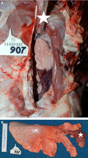
Figure 2.

Right lung with lobar consolidation (LS4) of the cranial aspect of the right cranial lobe. (A) In situ specimen. Carcass is in left lateral recumbency. The right 2nd rib (white star) is reflected dorsally exposing the combined 1st and 2nd intercostal space, revealing the cranial aspect of the right cranial lobe. (B) Right lung removed, showing consolidation of cranial aspect of cranial lobe (white star).
