Figure 3.

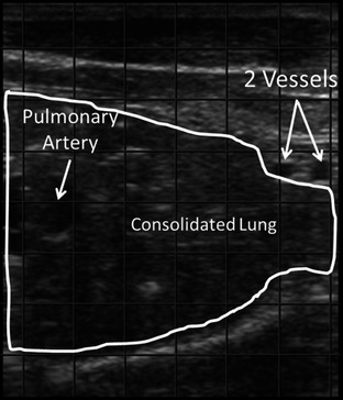
Ultrasonographic image of consolidated lung imaged from the right 1st intercostal space. Image orientation: left = dorsal, right = ventral, top = superficial, bottom = deep. The 2 blood vessels are the internal thoracic artery and vein.

Ultrasonographic image of consolidated lung imaged from the right 1st intercostal space. Image orientation: left = dorsal, right = ventral, top = superficial, bottom = deep. The 2 blood vessels are the internal thoracic artery and vein.