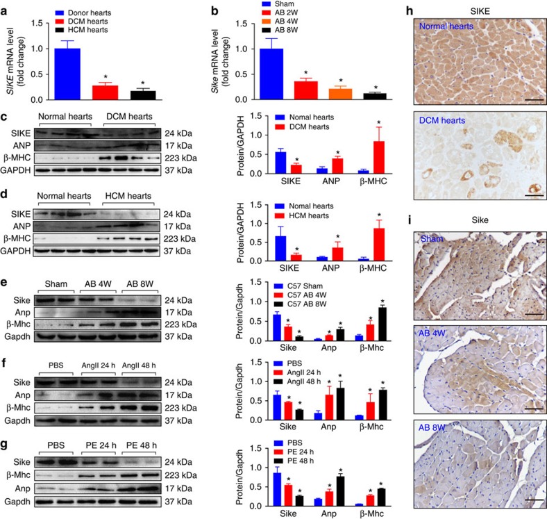
Figure 1. SIKE expression is downregulated in hypertrophic hearts.
(a) SIKE mRNA levels in normal, DCM and HCM human hearts. (b) Sike mRNA levels in the hearts of sham-operated controls and AB-induced cardiac hypertrophic mice at the indicated times. Values represent the fold changes relative to the sham controls, set as 1; the GAPDH mRNA level was used as the internal control in a and b. (c–g) Immunoblot and quantification of SIKE, ANP and β-MHC protein levels in normal and DCM human hearts (c), normal and HCM human hearts (d), control and 4- to 8-week AB-induced hypertrophic mouse hearts (e), and 1 μM Ang II-treated (f) or 100 μM PE-treated (g) neonatal rat cardiomyocytes (NRCMs) for the indicated times; n=4 samples or repeats/group; the GAPDH protein level was used for normalization. (h,i) Representative images of immunohistochemical staining of normal or DCM human heart sections (h) and control or hypertrophic mouse heart sections (i) with an antibody against SIKE; scale bars, 50 μm. *P<0.05 versus the corresponding controls (normal hearts, sham-operated mouse hearts or PBS-treated cardiomyocytes in a–g). Data are presented as the mean±s.d. from at least three independent experiments. For c and d, statistical analysis was carried out by Student's two-tailed t-test; for a,b and e–g statistical analysis was carried out by one-way analysis of variance.

