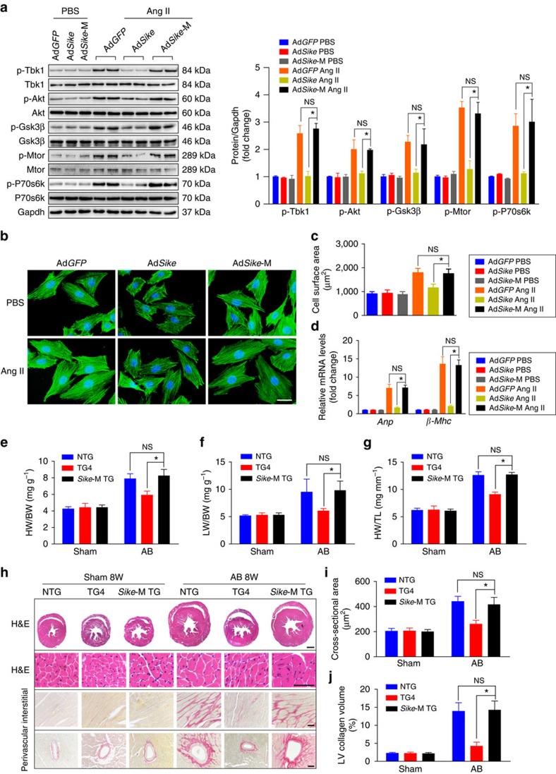
Figure 11. SIKE ameliorate cardiac remodelling dependent on SIKE-TBK1 interaction.
(a–d) NRCMs were infected with the indicated adenovirus, followed by PBS or Ang II treatment. Immunoblotting of the active levels of Tbk1 and Akt signalling components was performed (a); NRCMs were stained with an α-actinin antibody and DAPI to measure the average cell surface area (b,c; n≥50 cells per group, scale bar, 20 μm); the mRNA levels of the hypertrophic marker genes were compared in the indicated groups (d). (e–g) Comparison of the HW/BW (e), LW/BW (f) and HW/TL (g) ratios in different genotypic mice subjected to sham or AB surgery, n=10–13 mice per group. (h) Histological analyses of whole hearts (the first row; scale bar, 1,000 μm) and heart sections from the indicated groups stained with H&E (the second row; scale bar, 50 μm) or PSR (the third and fourth row; scale bars, 50 μm) 8 weeks after sham or AB surgery, n=6–8 mice per group. (i) Comparison of the cross-sectional area of cardiomyocytes, n≥100 cells per group. (j) Comparison of the LV collagen volume in the indicated groups, n≥40 fields per group. *P<0.05 compared between the two indicated groups; NS indicates no significance. Data are presented as the mean±s.d. from at least three independent experiments. Statistical analysis was carried out by one-way analysis of variance.

