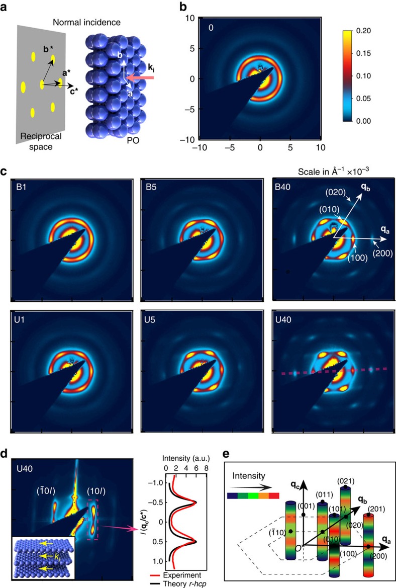
Figure 7. SAXS structure factor of BIOS-processed opals.
(a) Illustration of SAXS at normal incidence with incident wave vector ki, unit vectors a,b of hexagonal lattice, a*, b* and c* are corresponding reciprocal lattice unit vectors. (b,c) SAXS patterns of different samples at normal incidence, with qa,b along a*, b*, respectively. (d) Left, grazing incidence SAXS pattern (see inset geometry) showing scattering along selected rods on the red dashed line of sample U40 in c. Right, extracted scattering intensity along rod (10l) compared to scattering from r-hcp structure in theory. Separation between scattering peaks corresponds to layer spacing of 197 nm in real space (F=5.1 μm−1). (e) Intensity distributions along different rods, adjusting for the form factor. All SAXS images are (10 × 10) × 10−3 Å and on the same colour scale.

