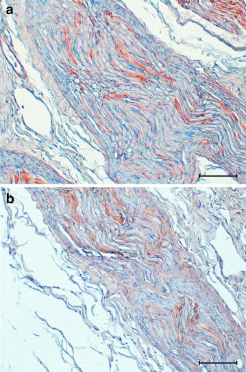
Fig. 4.

Photomicrographs of the immunohistochemical reactivity of the sciatic nerve. Sciatic nerve from NAD-affected dog (a) and healthy control dog (b). Longitudinal section of sciatic nerve shows weak linear immunohistochemical reactivity for GFAP both in affected (a) and healthy dog (b). Avidin–biotin-peroxidase complex method with Mayer’s hematoxylin counterstain. Bar = 100 µm
