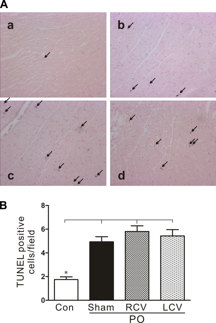
Fig. 8.
Moderate increases in the number of apoptotic myocytes with PO are unaffected by RCV or LCV therapies. A: representative terminal deoxynucleotide transferase-mediated nick-end labeling (TUNEL) in CardioTACS-stained sections of LV tissue from control (a) and PO-treated [PO + sham (b), PO + RCV (c), and PO + LCV (d)] hearts. Arrows indicate blue-stained nuclei, indicative of DNA fragmentation, a hallmark of apoptosis. B: quantification of apoptotic cells in experimental groups shown in A. *P < 0.05 vs. all PO.

