Fig. 1.
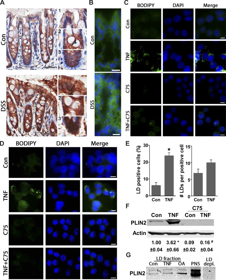
Increased lipid droplets (LDs) associated with colonic inflammation are mediated via TNF-dependent lipogenesis. A: colonic tissues from control and dextran sulfate sodium (DSS)-treated mice were immunohistostained for PLIN2 (n = 4, scale bar 50). Insets (1–3): enlarged areas of apical, intermediate, and basal portions of the colonic crypts. B: tissues from the same mice were stained with BODIPY 493/503 (n = 4, scale bar = 10 μm). C and D: human colonic NCM460 (nontransformed) and HT-29 (transformed) cells were stimulated with TNF (6 h) with or without C75 and stained for LDs (BODIPY 493/503) (representative images from 3 independent experiments, scale bar = 5 μm). E: graphs present the number of LD-positive cells and LDs per cell in HT-29 cells treated with TNF (6 h) (ImageJ quantification of BODIPY 493/503 stained, n = 300 cells, *P < 0.05 compared with Con). F: total protein from HT-29 cells treated with TNF (6 h) with or without C75 were immunoblotted for the LD coat protein Perilipin 2 (PLIN2) (n = 3, *P < 0.05, compared with Con, #P < 0.05, compared with TNF alone, ANOVA). G: LD fractions obtained from HT-29 control cells, TNF (24 h), and oleic acid (OA, 24 h) were immunoblotted for PLIN2. As a positive control, protein from postnuclear supernatant (PNS) was used, and as negative control LD depleted fraction (LD depl.) from control cells was used.

