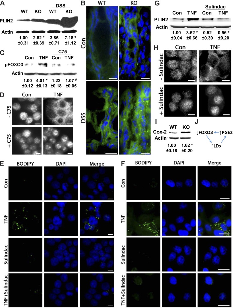
Fig. 3.
TNF-induced increases in LDs depend on prostaglandin E2 (PGE2) and FOXO3 interplay. A: protein from scraped colonic mucosa of wild-type (WT) and Foxo3-deficient knockout (KO) mice with or without DSS treatment were analyzed for PLIN2 expression by immunoblotting (n = 4, *P < 0.05, compared with Con, # P < 0.05, compared with KO alone, ANOVA). B: BODIPY (493/503) staining of colonic tissues from WT and Foxo3 KO mice treated with DSS (n = 4, scale bar = 10 μm). C: protein from HT-29 cells stimulated with TNF (0.5 h) with or without C75 was immunoblotted for pFOXO3 (corresponding densitometric analysis n = 3, *P < 0.05, compared with Con, #P < 0.05, compared with TNF alone, ANOVA). D: HT-29 cells treated with TNF (1 h) in the presence of C75 were immunofluorescently stained for FOXO3 (Alexa 488); representative images from 3 independent experiments, scale bar = 5 μm. E and F: NCM460 and HT-29 cells treated with TNF (6 h) in the presence of the PGE2 synthesis inhibitor sulindac were stained for LDs (BODIPY 493/503); representative images from 3 independent experiments, scale bar = 5 μm and 10 μm. G: immunoblotting and corresponding densitometric analysis of protein from HT-29 cells treated with TNF (6 h) with and without sulindac (n = 3, *P < 0.05, compared with Con, #P < 0.05, compared with TNF alone, ANOVA). H: HT-29 cells treated with TNF (1 h) with or without sulindac were immunofluorescently stained for FOXO3 (Alexa 488; representative images from 3 independent experiments, scale bar = 5 μm). I: protein from scraped colonic mucosa of WT and Foxo3 KO mice was immunoblotted for cyclooxygenase 2 (COX-2) (n = 3, *P < 0.05 compared with WT, t-test). J: proposed model of LD-FOXO3-PGE2 negative regulatory loop.

